Machine learning assistive two-dimensional ´molecular fingerprint´ of blood
Weng Kung Peng, a researcher from International Iberian Nanotechnology Laboratory (INL), Portugal, in collaboration with researchers from Singapore developed a new technique to map out two-dimensional ´molecular fingerprint´ from a single drop of blood (<1μL) using home-built NMR-based micro scanner.
“This highly time‒ and patient-specific information is akin to generating a unique QR-code for an individual that reflects his/her biological status at that point in time. This is one of the ´sci-fi´ moments when we realized that we may be able to identify a spectrum of diseases accurately based on the ´molecular fingerprint´ of the blood”, said Dr PENG, the leading author of this work. So far they have mapped out various physiological conditions (e.g., oxygenated/deoxygenated states) and confirmed the molecular fingerprint of several disease states (e.g., blood oxidation damages, hemoglobinopathies). Dr PENG is optimistic of leveraging on the technology to achieve breakthroughs in other diseases (e.g., malaria, diabetes mellitus and cancer). This is probably one of the most important milestones in the history of magnetic resonance after the development of contrast agent for magnetic resonance imaging (MRI), the author added.
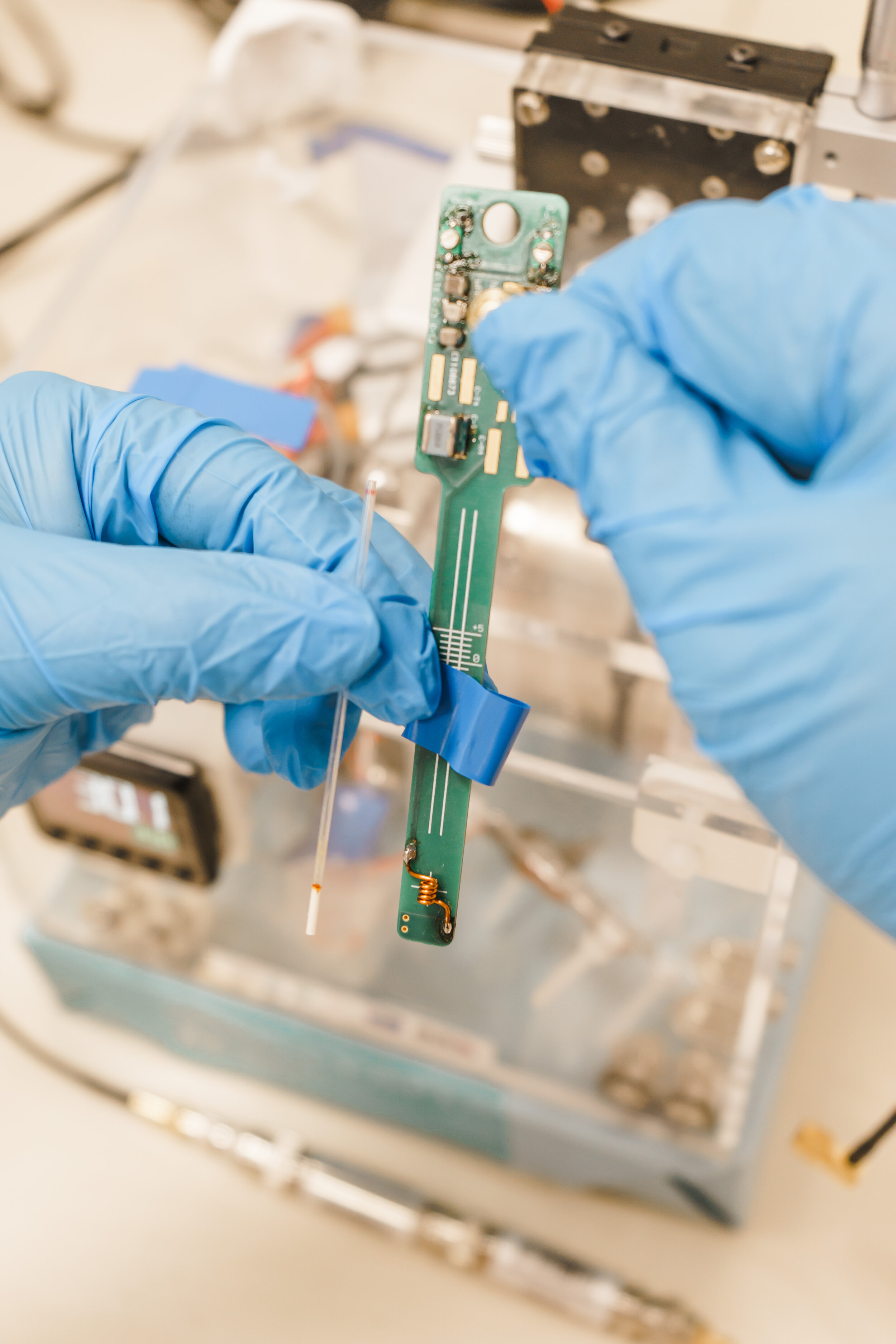
Magnetic resonance technology is always known to be physically large, sophisticated machines that cost millions. The team pivoted its research in the opposite direction and developed a low-cost, simplified version of the micro scanner, which is compact and portable. Instead of whole-body imaging (as in the case of MRI), the team focused on micro-volume imaging (a drop of blood). The team replaced the large and expensive items (e.g., magnetic chamber and gradient coils), with an inexpensive coin-sized magnet (aka fridge magnet) and hand-woven coil. In a surprising turn, smaller coil produced a much focused and hence higher power output. And this in return, removed the need for the bulky and expensive power amplifier.
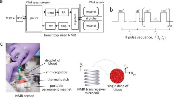
Fig. 1: Two-dimensional NMR T1-T2 correlational spectroscopy for molecular phenotyping of blood.
The NMR correlational spectroscopy is in the direct analogy of magnetic resonance imaging, which displays the images in the physical dimensions. In spectroscopy, however, the two-dimensional correlational maps represent complex signatures with complicated biological pathways. It took the team a while to decipher the complex data until Dr PENG chanced upon a machine learning lecture one day. Out of curiosity, the team fed a set of unlabelled NMR correlational maps taken from human subjects (with and without disease) into a machine learning algorithm, and it successfully clustered the data according to the type and severity of the disease. Machine learning (ML) is good at differentiating complex features and recognise the subtle characteristics which may not be obvious to the human eyes. The team found that ML is able to recognise an image much faster and accurate than a human being in a blinded test. This is probably one of the most important moment for ML after the winning of Google DeepMind against World champion Lee Sedol in Go match in 2016. We should not fear ML, instead, embrace and make full use of the symbiosis between Human-Machine (Learning) interactions. This is the beginning of a new era (in the making), where ML will soon be part of our life. If I have to make a comparison, it will be like witnessing the pre- and post-internet era, Dr PENG added.
“This work is game-changer for several reasons. Most of the disease markers are currently described in one dimension (high/low). Two-dimensional states allow the more complex representation of biological systems. Second, the NMR-based micro scanner developed here opens new avenues for non- or minimally-invasive in vitro diagnostic for patient care.” said Dr Tze Ping Loh, who is the Research Director (Laboratory Medicine) of the National University Hospital, Singapore, and one of the senior authors of this work.
A paper of the team´s breakthrough is published in the journal Communications Biology, a prestigious journal by Nature Publishing Group. You can find the publication here.